
09 – Endocrine System
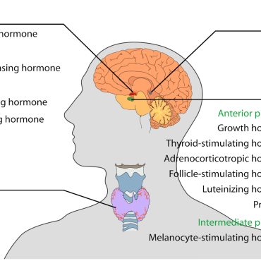
This episode provides a comprehensive review of the endocrine system, covering hormone types, signaling pathways, and feedback regulation. We tour the major endocrine glands and their hormones, and explain key […]
 play_arrow
 play_arrow
10 – Cardiovascular System 1 of 2 The Heart
Mark
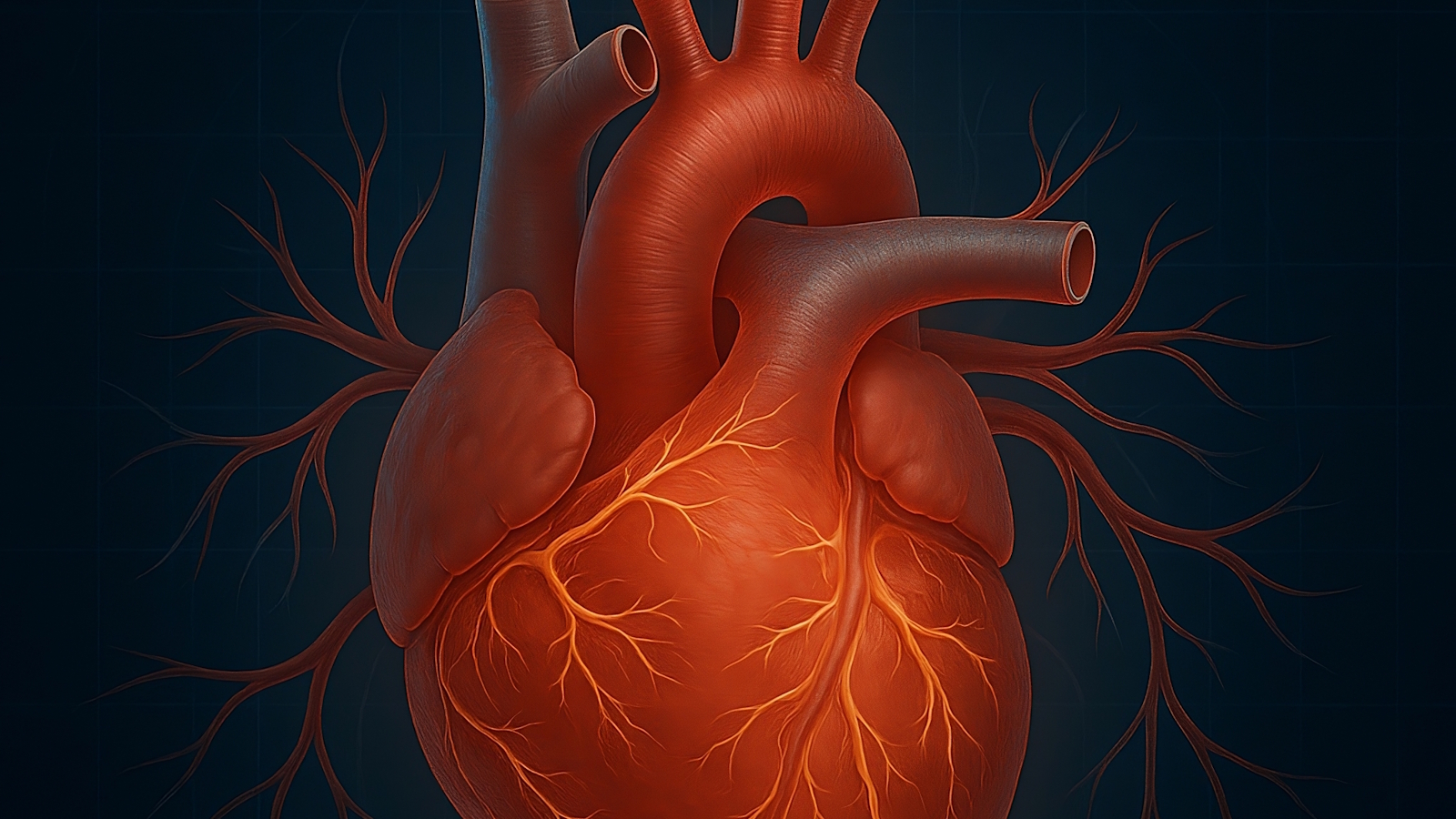
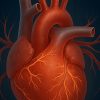
In this episode of the MCAT Prep Pod Biology Review, we take a deep dive into the heart’s role in the cardiovascular system, the body’s essential transport network. We begin by outlining its core functions: the transport of oxygen, nutrients, and hormones; the removal of waste; and its critical roles in regulating temperature, pH, and fluid balance.
We then explore the detailed anatomy of the heart, covering the three layers of the heart wall (endocardium, myocardium, epicardium), the four chambers, and the pathway of blood through the four critical valves. We provide helpful mnemonics like LAB RAT (Left Atrium Bicuspid, Right Atrium Tricuspid) and “Try NOT Pulling My Aorta” to remember the valve order. The discussion includes the heart’s own blood supply via the coronary arteries.
Next, we trace the complete pathway of blood through both the low-pressure pulmonary circuit and the high-pressure systemic circuit. This section clarifies the journey of deoxygenated blood to the lungs and oxygenated blood to the rest of the body, highlighting MCAT-favorite exceptions like the pulmonary artery and veins.
The episode then breaks down the cardiac cycle, explaining the phases of systole (contraction) and diastole (relaxation). We connect these mechanical events to the heart sounds (S1 and S2) and important cardiac volumes like stroke volume and ejection fraction. Key physiological concepts such as preload, afterload, and the Frank-Starling mechanism are explained in detail, along with the calculation for cardiac output.
Finally, we uncover the heart’s electrical conduction system, from the sinoatrial (SA) node’s role as the natural pacemaker to the AV node, bundle of His, and Purkinje fibers. We explain how pacemaker cells achieve autorhythmicity through “funny” channels and how the calcium plateau in ventricular myocytes prevents tetany. This leads to a clear explanation of the electrocardiogram (ECG), detailing what the P wave, QRS complex, and T wave represent in the heart’s electrical cycle.
This episode provides a comprehensive review of the endocrine system, covering hormone types, signaling pathways, and feedback regulation. We tour the major endocrine glands and their hormones, and explain key […]

Post comments (0)